CBCT Cone Beam Ann Arbor, MI
Dental cone beam computed tomography, or CBCT cone beam, is a type of X-ray equipment that uses a 3D cone beam and dental scans to provide dentists with a detailed, three-dimensional image of the mouth. Furthermore, patients do not have to suffer any pain or discomfort during the procedure. As oral surgeons, we can use a CBCT cone beam to take detailed photos of the bones, nerve pathways, soft tissues, and teeth with just a single scan.
CBCT cone beam treatment is available in Ann Arbor and the surrounding area. Great Lakes Oral Surgery uses this advance in dental technology to plan successful procedures. Call us today at (734) 961-4864 to schedule an appointment or learn more about our services.
Understanding CBCT Cone Beam
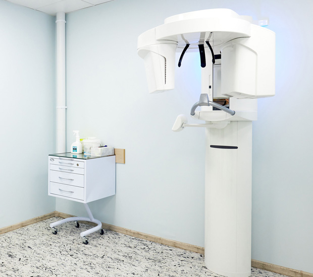
CBCT cone beam is a special type of X-ray machine utilized when regular dental or facial X-rays are insufficient. CBCT cone beams can generate 3D images of dental structures, soft tissues, nerve paths, and bone in the craniofacial region in a single scan using a special kind of technology. As such, CBCT cone beam images allow our oral surgeon to plan for more precise treatment.
While the CBCT cone beam is not the same as conventional CT, it can still be used to produce images similar to the ones made by traditional CT imaging. With a CBCT cone beam, an X-ray beam in the shape of a cone moves around the patient to produce multiple images, also known as views. Both CT scans and CBCT cone beam produce high-quality images, though CBCT cone beam utilizes a much smaller machine.
“CBCT cone beam is a special type of X-ray machine utilized when regular dental or facial X-rays are insufficient.”
How CBCT Cone Beam Works
CBCT cone beam scanners are square-shaped machines, including an upright chair for patients to sit on or a moveable table for patients to lie down during the examination. Scanners with a chair have a rotating C-arm, a device that intensifies X-ray images and contains an X-ray source and detector. Scanners with a table include a rotating gantry.
The C-arm or gantry will rotate around the head in a single 360-degree rotation, capturing multiple images from different angles during the examination. These images will then be reconstructed to create a single 3D image. The X-ray source and detector will be mounted on opposing sides of the revolving C-arm or gantry. They will rotate in unison, generating anywhere from 150 to 200 high-resolution 2D images. These are then digitally combined to form a 3D image, providing the oral surgeon with valuable information about the patient's oral and craniofacial health.
“CBCT cone beam scanners are square-shaped machines, including an upright chair for patients to sit or a moveable table for patients to lie down during the examination.”
CBCT Cone Beam Results
CBCT cone beam scanners render far more detailed images than traditional CT scanners. Consequently, as previously established, they are particularly useful when traditional X-rays are insufficient to provide necessary treatment information. For example, CBCT cone beams can evaluate the bony structures of the face, dentition, diseases of the jaw, nasal cavity, and sinuses.
CBCT cone beam technology can also be useful in diagnosing oral cancers and cysts and managing impacted teeth. According to one study, CBCT cone beam technology was found to have contributed significantly to the planning and successful surgical management of dentigerous cysts and associated impacted teeth. In addition, as cone beams take wide-range photos, they can capture in-depth areas that traditional scanners may miss.
“CBCT cone beam scanners render far more detailed images than traditional CT scanners.”
Check out what others are saying about our dental services on Yelp: CBCT Cone Beam in Ann Arbor, MI
After the CBCT Cone Beam Procedure
Once the CBCT cone beam procedure has been completed, the oral surgeon will have a clear, 3D image of the teeth, mouth, jaw, neck, ears, nose, and throat. It is possible to review this image immediately and get the best possible view of any area with features such as zooming and rotation. In addition, this is a painless procedure with no recovery time, and the patient will be able to return to their everyday activities immediately.
The practitioner will also write a report of the scan results, and the patient will be able to see their images. Finally, the surgeon and patient will go over the treatment plan together, during which time the patient can discuss their preferences for the treatment process and aftercare.
“Once the CBCT cone beam procedure has been completed, the oral surgeon will have a clear, 3D image of the teeth, mouth, jaw, neck, ears, nose, and throat.”
Questions Answered on This Page
Q. How do CBCT cone beam scanners work?
Q. What is the difference between CBCT cone beam scanners and traditional CT scanners?
Q. What happens after the CBCT cone beam procedure?
People Also Ask
Q. Do missing or crowded teeth affect dental implant candidacy?
Q. How is an oral surgeon different from a general dentist?
Q. What are the different types of sedation?
Q. What other common issues can prevent a person from being a dental implant candidate?
Frequently Asked Questions
Q. Will my insurance cover CBCT cone beam scans?
A. Some insurance companies may cover CBCT cone beam scans. However, this is not a guarantee, as all plans are different. Your best bet is to contact your insurance provider directly. Our oral surgeon can help you figure out what is covered in your plan.
Q. What kind of image is produced by a CBCT cone beam scan?
A. CBCT cone beam scanners take detailed images of the entire head. As teeth positioning affects the entire head, these images allow us to see the underlying bone structure and jawline. With this information, our oral surgeon can create the most accurate treatments possible. CBCT cone beam scanners are particularly beneficial for procedures around delicate areas. They are also useful for planning correctional procedures.
Q. How long does a CBCT cone beam scan take?
A. These scans are typically brief. For example, a CBCT cone beam scan only needs a singular rotation around the head. Once you are positioned, the scan itself will usually be over in less than 30 seconds.
Q. How long have oral surgeons been using CBCT cone beam technology?
A. According to the Food and Drug Administration (FDA), dental professionals have been using CBCT cone beam technology for over twenty years. Furthermore, dental scans are becoming increasingly common, thanks to their helpfulness in procedure planning and diagnosing complex conditions.
Q. How much radiation does a CBCT cone beam scan emit?
A. Though CBCT cone beam scans are still considered computed tomography (CT) scans, they emit considerably less radiation than conventional CT scans. However, it does emit more radiation than a traditional X-ray. Therefore, our oral surgeon will only recommend a CBCT cone beam scan if they judge the benefits greater than the risks.
Q. How should I prepare for a CBCT cone beam scan?
A. There is very little preparation necessary for a CBCT cone beam scan. Make sure to remove all loose or metallic objects before the exam. Stay still and do not swallow or talk during the scan.
Start Feeling Better – Visit Us Today
By visiting us as soon as possible, our team can help get you the professional treatment you need. Instead of waiting around and allowing the symptoms to get worse, we can provide you with treatment options.
Call Us Today
CBCT cone beam technology can give our oral surgeon the necessary information to personalize the best treatment plan for you. Our team at Great Lakes Oral Surgery in Ann Arbor can help. Call us today at 734-961-4864 to schedule an appointment or learn more about our services.
Helpful Related Links
- American Dental Association (ADA). Glossary of Dental Clinical Terms. 2024
- American Academy of Cosmetic Dentistry® (AACD). Home Page. 2024
- American Academy of Maxillofacial Prosthetics. American Academy of Maxillofacial Prosthetics. 2024
- American Association of Oral and Maxillofacial Surgeons. American Association of Oral and Maxillofacial Surgeons. 2024
- American College of Oral and Maxillofacial Surgery. American College of Oral and Maxillofacial Surgery. 2024
- National Cancer Institute (NCI). National Cancer Institute (NCI). 2024
- WebMD. WebMD’s Oral Care Guide. 2024
About our business and website security
- Great Lakes Oral Surgery was established in 2024.
- We accept the following payment methods: American Express, Cash, Check, Discover, MasterCard, and Visa
- We serve patients from the following counties: Washtenaw County
- We serve patients from the following cities: Ann Arbor, Ypsilanti, Saline, Dexter, and Chelsea
- Norton Safe Web. View Details
- Trend Micro Site Safety Center. View Details
Back to top of CBCT Cone Beam


